|
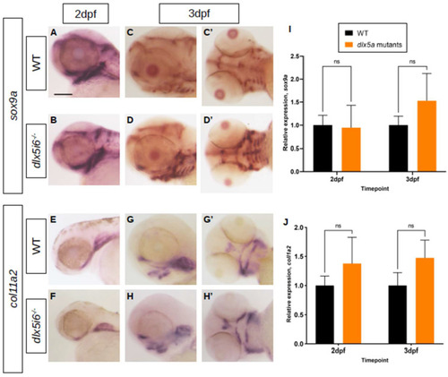
Expression of chondrocyte markers at 3 dpf in dlx5i6−/− larvae. (A–D) WISH for sox9a at 2 dpf (A,B) in WT and dlx5i6−/− larvae in the lateral position. (C,D) WISH for sox9a at 3 dpf in WT and dlx5i6−/− larvae in lateral (C,D) and ventral (C′,D′) positions, showing more staining for this marker in dlx5i6−/− mutants. (E,F) WISH for col11a2 in 2 dpf WT (E) and dlx5i6−/− larvae (F) in lateral position. (G,H) WISH for col11a2 in 3 dpf WT (G,G′) and mutant (H,H′) in lateral and ventral positions. Scale = 100 μm. (I,J) Relative normalized expression of sox9a (I) and col11a2 (J) at 2 dpf (p = 0.32; p = 0.16, respectively) and 3 dpf (p = 0.42; p = 0.24, respectively). ‘ns’: not significant.
|

