FIGURE

Figure 2

ID
ZDB-FIG-231002-173
Publication
Yu et al., 2023 - Loss of dlx5a/dlx6a Locus Alters Non-Canonical Wnt Signaling and Meckel's Cartilage Morphology
Other Figures
All Figure Page
Back to All Figure Page
Figure 2

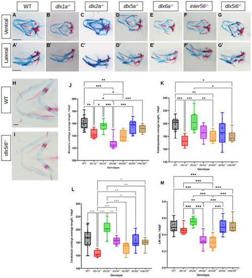
Alcian blue and alizarin red staining of 14 dpf dlx mutants reveal altered lengths of craniofacial structures. (AG) Histology stains were performed in at least 10 larvae for each genotype. Images were taken in ventral (AG) and lateral (A′G′) positions. Scale = 150 μm. (H,I) Flat mount of dlx5i6−/− (n = 5) and WT siblings (n = 6) in the ventral position to better show MC structure. Scale = 100 μm. (JL) Average lengths of MC (J), PQ (K) and CH (L) in 14 dpf animals were obtained following the same method as 5 dpf. At least 8 animals per genotype were measured. Results are reported as mean ± SEM. (M) l/w ratio in dlx5a−/− and dlx5i6−/− larvae continue to be smaller compared to WT. One-way ANOVA with Tukey’s multiple comparisons test was performed. * p < 0.05; ** p < 0.001; *** p < 0.0001.

Expression Data

Expression Detail
Antibody Labeling
Phenotype Data

Phenotype Detail
Acknowledgments
This image is the copyrighted work of the attributed author or publisher, and ZFIN has permission only to display this image to its users. Additional permissions should be obtained from the applicable author or publisher of the image. Full text @ Biomolecules