Fig 4
- ID
- ZDB-FIG-230916-107
- Publication
- Walker et al., 2023 - Target-selective vertebrate motor axon regeneration depends on interaction with glial cells at a peripheral nerve plexus
- Other Figures
- All Figure Page
- Back to All Figure Page
|
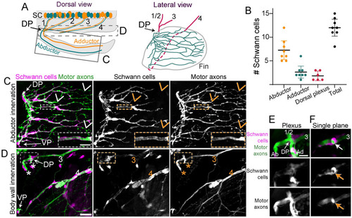
Schwann cells tightly associate with axons in the pectoral fin. (A) Schematic of a dorsal view of a larval zebrafish. In the SC, motor axons that sort at the DP to innervate the abductor (green) or adductor (orange) muscle are mixed within nerves 1–3. Nerve 4 axons sort at the VP (not labeled). The C and D boxes denote the regions included in the maximum projections shown in C and D. The lateral view shows the arrangement of nerves 1–4 in the body wall (magenta) and innervation in the fin (green) as it is shown in C and D. The dashed line indicates where nerve 4 grows in the body wall behind the fin. (B) Quantification of number of Schwann cells on the abductor or adductor muscle or associated with the plexus; |

