
Fig 2
- ID
- ZDB-FIG-230916-105
- Publication
- Walker et al., 2023 - Target-selective vertebrate motor axon regeneration depends on interaction with glial cells at a peripheral nerve plexus
- Other Figures
- All Figure Page
- Back to All Figure Page
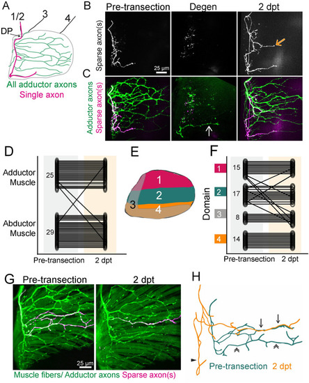
Target-selective regeneration of motor axons. (A) Schematic of a lateral view of pectoral fin abductor muscle motor innervation. An example trajectory of a single axon is shown in magenta. DP labels dorsal plexus, motor nerves in the body wall are labeled 1–4. (B, C) A timecourse of sparsely labeled axon(s) showing the innervation pattern pre-transection, after axon degeneration, and at 2 dpt. Sparsely labeled axons are labeled in white (B) and magenta (C) and all adductor motor axons are labeled in green (C). The white arrow points to a fascicle that did not degenerate and the orange arrow points to ectopic growth during regeneration. (D) Quantification of the muscle localization of sparsely labeled axons pre-transection compared to where these labeled axons innervated after regeneration. Small numbers on pre-transection data represent |