|
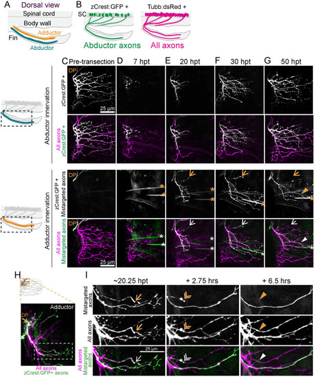
Mistargeted axons are selectively retracted. (A) Dorsal view schematic labeling abductor and adductor musculature of the fin. (B) Schematic of zCrest:GFP+ motor neurons, which project to the abductor muscle and Tubb:dsRed+ motor neurons, which project to both abductor and adductor muscles. zCrest:GFP+ motor neurons are also labeled with Tubb:dsRed. (C–G) Timecourse of regeneration of innervation on abductor (top) and adductor (bottom) musculature. Schematics on the left show the area included in maximum projections. Arrows point to mistargeted axons that will retract. Asterisks indicate muscle fibers also labeled by zCrest:GFP. DP labels dorsal plexus. (C) zCrest:GFP+ axons are not present on the adductor muscle before axon transection. (D) At 7 hpt, axons have fragmented. (E) At 20 hpt, regenerating zCrest:GFP+ axons are present on both the abductor and adductor muscle. (F, G) At 30 and 50 hpt, some zCrest:GFP+ mistargeted axons have retracted (arrow), whereas other mistargeted axons persist (triangle). (H) Maximum projection of axon regeneration onto adductor muscle at 20.25 hpt. The boxed region is expanded in I. zCrest:GFP+ axons are mistargeted onto adductor muscle. (I) Images from timelapse imaging of axon regeneration onto adductor muscle. zCrest:GFP labels mistargeted axons, whereas axons that are only magenta (labeled with Tubb:dsRed) are correctly targeted to adductor muscle. Example of a mistargeted axon that is present at 20.25 hpt (arrow), retracting at +2.75 h (double arrowhead) and gone by +6.5 h (filled triangle). DP, dorsal plexus; GFP, green fluorescent protein; hpt, hour post transection.
|