
Fig. 4
- ID
- ZDB-FIG-230904-29
- Publication
- Huljev et al., 2023 - A hydraulic feedback loop between mesendoderm cell migration and interstitial fluid relocalization promotes embryonic axis formation in zebrafish
- Other Figures
- All Figure Page
- Back to All Figure Page
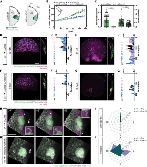
Figure 4. Prechordal plate migration is sufficient for interstitial fluid relocalization (A) Schematic of interstitial fluid (IF) distribution at 6 h post fertilization (hpf) (onset of prechordal plate [ppl] internalization in wild-type [WT] embryos) in MZoep gsc::caax-EGFP embryos containing transplanted ppl cells; D, dorsal and S, sagittal views; EVL, enveloping layer; DC, deep cell; YSL, yolk syncytial layer. (B) Distance of the ppl transplant leading edge from the germ margin as a function of developmental time (6 hpf, corresponding to 0′) in control (green, n = 6) and ouabain (O)-treated (blue, n = 9) MZoep;gsc::caax-EGFP host embryos containing untreated ppl cells, and control (light green, n = 4) and ouabain-treated (light blue, n = 2) MZoep;gsc::caax-EGFP host embryos containing ouabain-treated ppl cells; N, number of independent embryo replicates; schematic of distance measurements in upper left corner; mean ± SEM. (C) Number of lamellipodia and filopodia per time frame, normalized for the number of cells, in ppl cells taken from gsc::caax-EGFP donor embryos injected with LifeAct-RFP mRNA (50 pg) and transplanted into control (green, n = 3) and ouabain-treated (blue, n = 3) MZoep;gsc::caax-EGFP host embryos; individually plotted values represent protrusion numbers per minute and cell; mean ± SEM; Mann-Whitney test. (D–G) Maximum intensity projections (D, dorsal view) and single cross-sections (S, sagittal view) of MZoep (D and E) and ouabain-treated MZoep (F and G) gsc::caax-EGFP embryos containing transplanted ppl cells at 6 (D and F) and 8 hpf (E and G); for control embryos see Figures 1B and 1C; dashed white lines outline the ppl transplant; dashed blue lines outline the IF at the EVL-DC boundary (S, sagittal view); dashed pink lines outline the IF at the YSL-DC boundary (S, sagittal view); scale bars, 100 μm. (D′–G′) IF distribution profiles along the animal-vegetal (AV) axis relative to the position of ppl transplant, as indicated by gsc::caax-EGFP expression, in MZoep (D′ and E′) and ouabain-treated MZoep (F′ and G′) gsc::caax-EGFP embryos containing transplanted ppl cells at 6 (D′ and F′) and 8 hpf (E′ and G′); multi-color curves represent average values of n = 3 independent embryo replicates of the position of ppl/pam (black lines) and IF distribution at the EVL-DC (blue lines) and YSL-DC (pink lines) boundaries. (H) High resolution images of ppl cells taken from gsc::caax-EGFP donor embryos injected with LifeAct-RFP mRNA (50 pg) and transplanted into control and ouabain-treated MZoep;gsc::caax-EGFP host embryos injected with fluorescent dextran to label the IF at different times after transplantation (37′, 43′, and 59′, with 0′ corresponding to 6 hpf); arrowheads/inserts show the extension of an exemplary lamellipodium toward contact-free, IF-filled space; scale bars, 50 μm. (I,I') Orientation of lamellipodia (I) and filopodia (I′) per time frame, normalized for the number of ppl cells, taken from gsc::caax-EGFP donor embryos injected with LifeAct-RFP mRNA (50 pg) and transplanted into control (green, n = 3) and ouabain-treated (blue, n = 3) MZoep;gsc::caax-EGFP host embryos injected with fluorescent dextran to label the IF. n, number of independent embryo replicates. See also Figure S4 and Video S3. |
Reprinted from Developmental Cell, 58(7), Huljev, K., Shamipour, S., Pinheiro, D., Preusser, F., Steccari, I., Sommer, C.M., Naik, S., Heisenberg, C.P., A hydraulic feedback loop between mesendoderm cell migration and interstitial fluid relocalization promotes embryonic axis formation in zebrafish, 582-596.e7, Copyright (2023) with permission from Elsevier. Full text @ Dev. Cell