
Fig. 3
- ID
- ZDB-FIG-230904-28
- Publication
- Huljev et al., 2023 - A hydraulic feedback loop between mesendoderm cell migration and interstitial fluid relocalization promotes embryonic axis formation in zebrafish
- Other Figures
- All Figure Page
- Back to All Figure Page
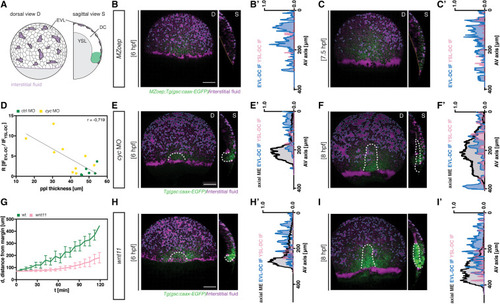
Figure 3. Axial mesendoderm internalization is required for efficient interstitial fluid relocalization (A) Schematic of interstitial fluid (IF) distribution at 6 h post fertilization (hpf) (onset of prechordal plate [ppl] internalization in wild-type [WT] embryos) in MZoep;gsc::caax-EGFP embryos; D, dorsal and S, sagittal views; EVL, enveloping layer; DC, deep cell; YSL, yolk syncytial layer. (B, C, E, F, H, and I) Maximum intensity projections (D, dorsal view) and single cross-sections (S, sagittal view) of MZoep (B and C), cyc morphant (MO, 4 ng) (E and F), and slb/wnt11f2 (H and I) gsc::caax-EGFP embryos at 6 hpf (B, E, and H) and 8 hpf (C, F, and I); for control embryos see (Figures 1B and 1C); dashed orange lines outline the YSL-DC boundary (S, sagittal view) in MZoep embryos; dashed white lines outline the ppl and, due to the leaky nature of this transgenic reporter line, also the posterior axial mesendoderm (pam); dashed blue lines outline the IF at the EVL-DC boundary; dashed pink lines outline the IF at the YSL-DC boundary (S, sagittal view); scale bars, 100 μm. (B′, C′, E′, F′, H′, and I′) IF distribution profiles along the animal-vegetal (AV) axis in MZoep mutant (B′ and C′), cyc MO (E′ and F′), and slb/wnt11f2 (H′ and I′) embryos relative to the position of ppl/pam, as indicated by gsc::caax-EGFP expression at 6 hpf (B′, E′, and H′) and 8 hpf (C′, F′, and I′); multi-color curves represent average values of n = 3 independent embryo replicates of the position of ppl/pam (black lines) and IF distribution at the EVL-DC (blue lines) and YSL-DC (pink lines) boundaries. (D) Correlation between IF relocalization and ppl thickness in control (green, n = 5) and cyc MO embryos (yellow, n = 4). Pearson correlation coefficient (r) is shown. (G) Distance of the ppl transplant leading edge from the germ ring margin as a function of developmental time (6 hpf, corresponding to 0′) in WT (green, n = 10) and slb/wnt11f2 (pink, n = 11) gsc::caax-EGFP embryos; mean ± SEM. n, number of independent embryo replicates. See also Figure S4. |
Reprinted from Developmental Cell, 58(7), Huljev, K., Shamipour, S., Pinheiro, D., Preusser, F., Steccari, I., Sommer, C.M., Naik, S., Heisenberg, C.P., A hydraulic feedback loop between mesendoderm cell migration and interstitial fluid relocalization promotes embryonic axis formation in zebrafish, 582-596.e7, Copyright (2023) with permission from Elsevier. Full text @ Dev. Cell