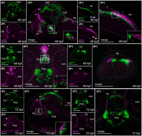
Fluorescence in situ hybridization confirms minimal co-localization of gsx1 and gsx2 during late embryonic and early larval development. (Ai-Avi) Lateral views showing expression of gsx1 and gsx2 at 48 hpf. (Bi-Bii) Ventral views showing expression of gsx1 and gsx2 at 48 hpf. (Biv-Bvi) Cross-sectional view taken at the dashed line in Avi. (Ci-Ciii) Lateral view showing gsx1 and gsx2 expression at 72 hpf. (Civ-Cvi) Ventral view showing gsx1 and gsx2 expression at 72 hpf. Lateral views were taken at ×20 and ventral/Cross-sectional views were taken at ×40, all with anterior facing left. All were pseudocolored using FIJI ImageJ and scale bars represent 100 μm. For lateral views, eyes were dissected off. Main images are max z-projections and insets are single z-stack slices zoomed into the boxed region shown in the main image. CeP, cerebellar plate; Di, diencephalon; Hb, hindbrain; Hyp, hypothalamus; Mb, midbrain; OB, olfactory bulb; P, pallium; Po, preoptic area; Pr, pretectum; Sc, spinal cord; Tel, telencephalon; TeO, optic tectum
|
