
FIGURE
Fig. 6
Fig. 6
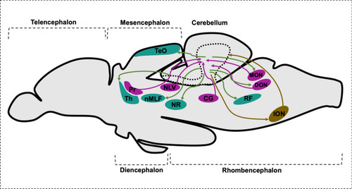
Schematic drawing of sagittal section of brain representing cerebellar afferents and canonical efferent population. CG central gray, DON descending octaval nucleus, ION inferior olive nuclei, MON medial octavolateralis nucleus, NLV nucleus lateralis valvulae, nMLF nucleus of the medial longitudinal fascicle, NR red nucleus, Pr pretectal nuclei, RF reticular formation, TeO optic tectum, Th thalamus |
Expression Data
Expression Detail
Antibody Labeling
Phenotype Data
Phenotype Detail
Acknowledgments
This image is the copyrighted work of the attributed author or publisher, and
ZFIN has permission only to display this image to its users.
Additional permissions should be obtained from the applicable author or publisher of the image.
Full text @ Cell. Mol. Life Sci.