
Fig. 2
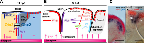
Molecular processes leading to the establishment of a patterned cerebellar primordium in zebrafish. a Schematic drawing of the establishment of the midbrain hindbrain boundary (MHB), a key secondary organizer tissue involved in inducing the cerebellar primordium in the dorso-anterior hindbrain. These processes are already initiated during mid-gastrulation stages, but gene expression is maintained in 14hpf embryos, where it can be assigned well to embryonic neuroanatomical structures. b At 24hpf, the cerebellar primordium is well established and patterned including the two major germinal zones, the upper rhombic lip (URL) and the ventricular zone (VZ) that give rise to all neurons in the adult cerebellum. (c Provided by Andreas Babaryka) Overlay of transmitted light images with results from double in situ hybridization at 36hpf against the mRNA of atoh1a (red) and ptf1a (blue) as molecular markers for these two juxtaposed proliferation zones. Transverse (left) and sagittal (right) vibratome sections through the developing cerebellum are displayed, scale bar. 50 µm. cb cerebellum, MHB midbrain-hindbrain boundary, rh rhombencephalon |