|
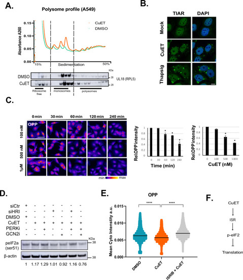
CuET rapidly blocks protein synthesis. A Upper panel: Polysome profiling of A549 epithelial cells following treatment with CuET for 1 h, lower panel: immunoblotting of the ribosomal protein RPL5 (uL18) in polysome profile fractions of A549 under the same experimental conditions. B Representative IF images of TIAR-containing stress granules after CuET treatment of A549 cells. The UPR inductor thapsigargin was used as a positive control. scale bar = 10 μm. C O-propargyl-puromycin (OPP) incorporation followed by high-content microscopy for the quantitation of translation rates in A549 cells treated with CuET or DMSO. 750–1500 cells were analyzed per experiment (data are shown as mean ± SD, n = 3 biological replicates, *p < 0.05) scale bar: 50 µm. D Immunoblotting of phosphorylated eIF2a (ser51) following treatment of A549 with CuET (2 h) +/–PERK inhibitor combined with siRNA against HRI or chemical inhibition of GCN2. Numbers below the blot indicate the signal ratio p-eIF2a/β-actin. E OPP incorporation followed by high-content microscopy for the quantitation of translation rates in A549 cells treated with CuET (2 h) +/− the ISR inhibitor ISRIB. 1000–2000 cells were analyzed per experiment (data shown as mean ± SD, n = 3 biological replicates, ****p < 0.001). F Schematic model connecting CuET treatment and translation.
|