|
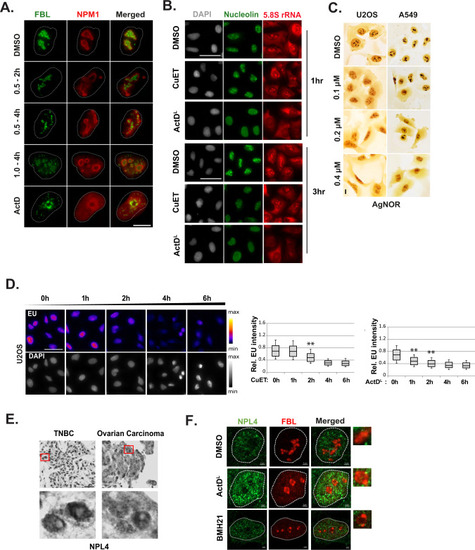
CuET alters the nucleolar morphology. A Representative IF images of A549 nuclei treated with CuET or ActDL for the indicated time points. Fibrillarin (FBL) or nucleophosmin (NPM1) were used as nucleolar markers. Scale bar: 5 μM. B Representative IF images of nucleolin and 5.8S rRNA levels in A549 cells treated with CuET or ActDL for the indicated time points. Scale bar: 50 µm. C AgNOR staining of U2OS or A549 cells following a four-hour treatment of increasing CuET doses. Scale bar: 10 μm. D Ethylene uridine (EU) levels were calculated following IF and high content imaging of U2OS treated with 1 μM CuET for the indicated time points. 750–1500 cells were analyzed per experiment (data are shown as mean ± SD, n = 3 biological replicates, **p < 0.01). Scale bar: 50 µm. E Detection of NPL4 protein levels with immunocytochemistry in samples from patients with triple-negative breast cancer (TNBC) or ovarian carcinoma. Regions in red squares are presented magnified in the bottom panel. Scale bar: 100 μM. F Representative IF images of nucleolar structure in U2OS treated with ActDL or BMH-21 as nucleolar stress inducers. Fibrillarin (FBL) was used as a nucleolar marker. Insets depict magnifications of the regions designated in squares. Scale bar: 2 μM.
|