|
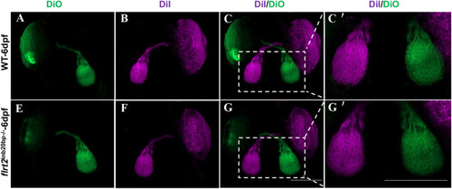
flrt2-KO zebrafish showed normal optic nerve projection. RGC axon projection in zebrafish at 6 dpf. Above: RGC axon projection in WT larvae, below: RGC axon projection in flrt2−/− mutant larvae. Retinotopic anterograde RGC axon labeling using DiI and DiO. n=20/each group. Scale bar: 200 μm. (A-G) LSM 980 with Airyscan was used: objective: plan-apochromat 10×/0.45 M27; scaling (per pixel): 0.829 µm×0.829 µm×8.790 µm; image size (pixel): 1024×1024; effective NA: 0.45; depth of focus: 5.43 µm. (C′,G′) LSM 980 with Airyscan was used: objective: plan-apochromat 20×/0.8 M27; scaling (per pixel): 0.414 µm×0.414 µm×3.760 µm; image size (pixel): 1024×1024; effective NA: 0.8; depth of focus: 1.72 µm.
|