|
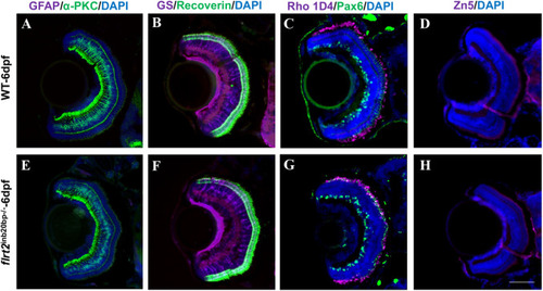
Retinal cells fate determination in flrt2-KO zebrafish. Immunofluorescence staining with retinal neural markers: (A,E) co-labelling with anti-GFAP antibodies (for glial cells, magenta) and anti-α-PKC antibodies (for bipolar cells, green). (B,F) Co-labelling with anti-GS antibodies (for Müller cells, magenta) and anti-Recoverin antibodies (for photoreceptor cells, green). (C,G) Co-labelling with anti-Rho 1D4 antibodies (for long double-cone outer segments, magenta) and anti-Pax6 antibodies (for ganglion and amacrine precursor cells, green). (D,H) Labelling with anti-Zn5 antibodies (for mature RGCs, magenta) in WT and flrt2-KO larvae retinas. Blue, DAPI staining of the nuclei. n=20/each group. Scale bar: 50 μm. ISM 710 was used: objective: LD plan-neofluar 20×/0.4 Korr M27; scaling (per pixel): 0.497 µm×0.497 µm; image size (pixel): 1388×1040; effective NA: 0.4; depth of focus: 8.02 µm.
|