|
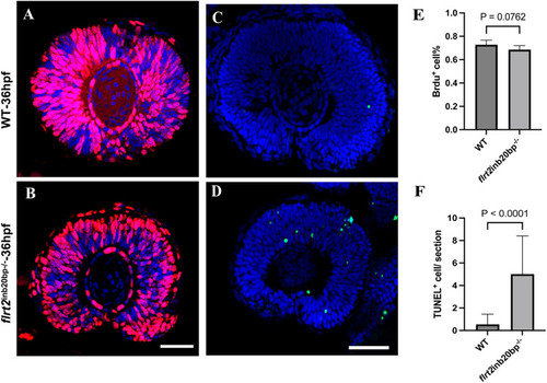
Retinal progenitor cell proliferation was not affected by the flrt-KO. (A,B) Images of 36 hpf zebrafish eye sagittal sections stained with anti-BrdU antibody (red). (C) Quantification of BrdU+ cells/section in WT and flrt2-KO retinas at 36 hpf; t=1.977, P=0.0762. Scale bars: 50 µm (n=6/each group). (D,E) TUNEL staining (green) of 36 hpf zebrafish eye sagittal sections. (F) Quantification of TUNEL+ cells/section in WT and flrt2-KO retinas at 36 hpf; t=4.911, P<0.0001. Scale bars: 50 µm (n=15/each group). The solid bars represent the means±standard deviations. LSM 980 with Airyscan was used: objective: plan-apochromat 20×/0.8 M27; scaling (per pixel): 0.414 µm×0.414 µm; image size (pixel): 1024×1024; effective NA: 0.8.
|