|
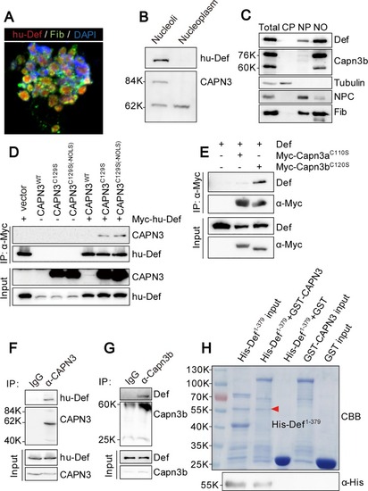
Def directly interacts with CAPN3, and they form a complex in the nucleolus. (A) Co-immunostaining of the endogenous hu-Def (red), Fib (green), and DAPI (blue) in the isolated nucleoli from the cultured HepG2 cells. (B) Western blot of the endogenous hu-Def and CAPN3 in the nucleoplasmic and nucleolar fractions from HepG2 cells. (C) Western blot of the endogenous Def, Capn3b, Tubulin, NPC, and Fib in different fractions as indicated isolated from the adult wild-type zebrafish liver. CP, cytoplasm; NP, nucleoplasm; NO, nucleoli; NPC, nuclear pore complex. (D) Co-IP of Myc-hu-Def and CAPN3. 293T cells were co-transfected with Myc-hu-Def and wild-type CAPN3 or mutant CAPN3 (CAPN3C129S and CAPN3C129S-ΔNOLS) plasmids. Total protein was extracted at 72 h and incubated with Myc-beads. Antibodies against CAPN3 and hu-Def were used in western blotting. (E) Co-IP of zebrafish Def and Myc-Capn3aC110S or Myc-Capn3bC120S. 293T cells were co-transfected with zebrafish def and Myc-Capn3aC110S or Myc-Capn3bC120S plasmids, respectively. Antibodies against zebrafish Def and the Myc-tag were used in western blotting. (F) Co-IP of the endogenous hu-Def and CAPN3 in HepG2 cells. Total protein extract was incubated with protein A/G agarose beads conjugated with anti-CAPN3 goat polyclonal antibody (COP-080049). CAPN3 was detected by a rabbit polyclonal antibody (No. 38963). (G) Co-IP of the endogenous Def and Capn3b in the isolated adult zebrafish liver nulceoli. Nucleolar protein extract was incubated with protein A/G agarose beads conjugated with anti-Capn3b antibody. (H) Def directly interacts with CAPN3. GST-pulldown was performed by incubating the purified His-Def1-379 with the purified GST, or GST-tagged wild-type CAPN3 immobilised on GST resin. Eluted proteins were stained either with Coomassie blue (CBB) (upper panel) or western blotting using an antibody against the His-tag (lower panel).
|