
Figure 5
- ID
- ZDB-FIG-230428-18
- Publication
- Frantz et al., 2023 - Pigment cell progenitor heterogeneity and reiteration of developmental signaling underlie melanocyte regeneration in zebrafish
- Other Figures
- All Figure Page
- Back to All Figure Page
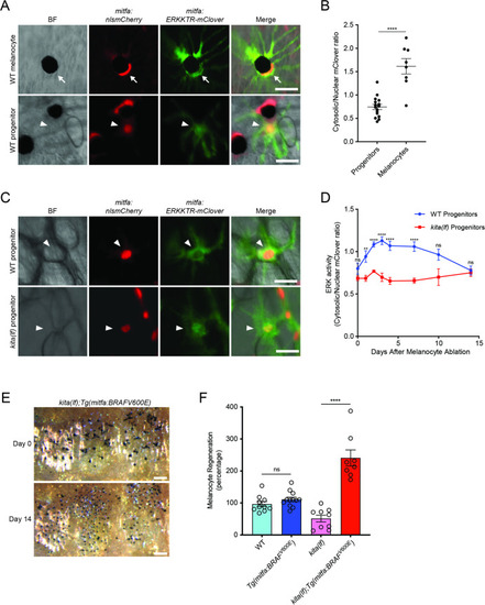
(A) Images of ERKKTR-mClover localization in a representative mature melanocyte (top, arrow) and progenitor (bottom, arrowhead) in uninjured wild-type animals. Scale bar = 30 µm. (B) Quantification of ERK activity in progenitors and melanocytes based on ERKKTR-mClover localization. Mean ± standard error of the mean (SEM) is shown; progenitors n = 16, melanocytes n = 8. (C) Images 3 days post-ablation of ERKKTR-mClover location in representative progenitors (arrowheads) in wild-type (top) and kita(lf) (bottom) animals. Scale bar = 30 µm. (D) Quantification of ERK activity in progenitors prior to and during melanocyte regeneration. For each data point, the average cytosolic/nuclear ratio of at least 6 cells ± SEM is shown. (E) Brightfield images of the melanocyte stripe before (top) and after (bottom) regeneration in kita(lf); Tg(mitfa:BRAFV600E) mutants. Scale bar = 200 µm. (F) Quantification of melanocyte regeneration in kita(lf);Tg(mitfa:BRAFV600E) and control animals. Mean percentage ± SEM is shown; wild-type n = 10, Tg(mitfa:BRAFV600E) = 12, kita(lf) = 9, kita(lf);Tg(mitfa:BRAFV600E) = 8 fish. p values calculated by Student’s t-test, **p < 0.01, ****p < 0.0001; ns, not significant.
|