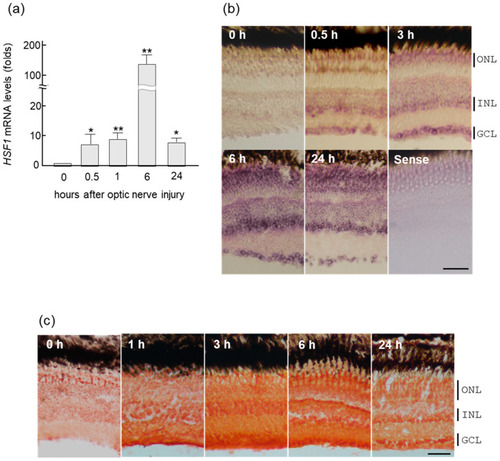
Upregulation of HSF1 (heat shock factor 1) mRNA in zebrafish retina after ONI (optic nerve injury). (a) HSF1 mRNA expression levels after ONI were determined using quantitative real-time PCR. (b) In situ hybridization of HSF1 in the zebrafish retina after nerve injury. HSF1 mRNA started to increase in the retina for 0.5 h and peaked at 6 h after ONI. Its localization was first seen in the GCLs (ganglion cell layers) and after the INLs (inner nuclear layers). Then, these signals spread to all nuclear layers including the ONLs (outer nuclear layers) at 6 h and slightly decreased at 24 h after ONL. (c) Immunohistochemical staining of HSF1 in the zebrafish retina after ONI. Significant immunostaining peaked at 3 to 6 h in all nuclear layers after ONI. Data are expressed as the mean ± SEM of five independent experiments and analyzed by one-way ANOVA, followed by Scheffe’s multiple comparisons. Statistical significance was set at * p < 0.05 or ** p < 0.01. Scale bar = 50 μm.