
Figure 4
- ID
- ZDB-FIG-230228-313
- Publication
- Sugitani et al., 2023 - Specific Activation of Yamanaka Factors via HSF1 Signaling in the Early Stage of Zebrafish Optic Nerve Regeneration
- Other Figures
- All Figure Page
- Back to All Figure Page
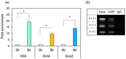
ChIP-enriched DNA was prepared using preimmune serum (IgG) or anti-HSF1 antibody from the control (0 h) or damaged zebrafish retina after ONI (6 h). (a) The immunoprecipitated DNA of Klf4, Oct4, and Sox2 were analyzed by real-time PCR. Each ChIP signal was divided by the no-antibody signals (IgG), representing the ChIP signals as the fold increase in signals relative to the background signals. (b) Gel electrophoresis image using the ChIP samples. The input was used as an internal positive control for the ChIP assay. Five to six experiments were repeated with different retinas under each experimental condition. Data are expressed as the mean ± SEM of independent experiments and analyzed by one-way ANOVA, followed by Scheffe’s multiple comparisons. Statistical significance was set at * p < 0.05. |