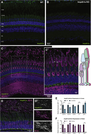
Impdh1a_tvX1 is expressed exclusively in rods and cones.A, representative IHC images showing Impdh1a staining of WT and (B) KO adult zebrafish retina at 11:00 AM using the C-terminal antibody. C, Impdh1a (green) and MTCO1 (magenta) immunostaining of WT-pigmented retina. The nuclei are blue. C′, magnified section of image in (C) showing Impdh1a filament localization in cones. The arrows indicate where Impdh1a filaments appear to form in the OS. Note the tiering of photoreceptors normal in adult zebrafish retina. D, representative Impdh1a staining (green) and 4C12 (magenta) of bleached retina showing the localization of Impdh1a with known zebrafish rod marker. The nuclei are stained blue. D′, magnified section of (D) showing overlap of Impdh1a with 4C12 (rod) staining. E, qPCR quantification of impdh1a, 1b, and 2 transcripts in coneless retina and (F) qPCR analysis of relative impdh1a, 1b, and 2 expression in rodless retina. es1 and gnat1 are cone and rod specific genes, respectively. N = 3 animals and the error bars are standard error. IHC, immunohistochemistry; impdh, inosine monophosphate dehydrogenase. ns p > 0.05, ∗ p ≤ 0.05 , ∗∗ p ≤ 0.01, ∗∗∗ p ≤ 0.001.