
Fig. 2
- ID
- ZDB-FIG-230215-9
- Publication
- Cleghorn et al., 2021 - A highly conserved zebrafish IMPDH retinal isoform produces the majority of guanine and forms dynamic protein filaments in photoreceptor cells
- Other Figures
- All Figure Page
- Back to All Figure Page
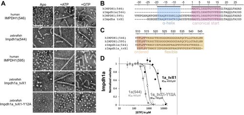
Structural and functional conservation between human and zebrafish IMPDH1.A, negative stain EM of purified human retinal variants IMPDH1(546) & IMPDH1(595) and zebrafish Impdh1a (544) & Impdh1a_tvX1. The scale bar represents 100 nm. B and C, sequence alignment of human retinal variants IMPDH1(546) and IMPDH1(595), zebrafish Impdh1a (544) {"type":"entrez-nucleotide","attrs":{"text":"NM_001002177.1","term_id":"50345007","term_text":"NM_001002177.1"}}NM_001002177.1, and zebrafish retinal variant Impdh1a_tvX1 ({"type":"entrez-nucleotide","attrs":{"text":"XM_005159007.4","term_id":"1207134073","term_text":"XM_005159007.4"}}XM_005159007.4). B, conserved N-terminal alpha-helix in blue and gene beginning in pink. C, residues in orange (ordered) are resolved in cryo-EM structures of the human protein (9), remainder of C-terminus in yellow is flexible and not resolved in structures (9). D, GTP-inhibition curves of zebrafish Impdh1a (544) (solid gray line), Impdh1a_tvX1 (solid black line), and nonassembly Y12A protein (dashed black line). Each data point represents a triplicate and the error bars are standard deviation. impdh, inosine monophosphate dehydrogenase; tv, transcript variant. |