
Fig. 6
- ID
- ZDB-FIG-230129-6
- Publication
- Wu et al., 2021 - Iterative tomography with digital adaptive optics permits hour-long intravital observation of 3D subcellular dynamics at millisecond scale
- Other Figures
- All Figure Page
- Back to All Figure Page
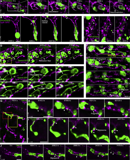
Figure 6. Membrane dynamics during tumor cell migration in zebrafish and mice (A) MIPs showing the vasodilation process before the tumor cell (MDA-MB-231, green, PH-mCherry) migrated and the vasoconstriction after the tumor cell was washed away by blood flow in zebrafish larvae. The contour profiles of the vessel are marked by dashed lines. (B) A tumor cell split a large vesicle when trapped in small-bore vessels (magenta, Tg(fli:GFP)) in zebrafish larvae (Video S5, part I). (C) Various vesicles flowing in the zebrafish vessel (Video S5, part II). (D and E) Formation of retraction fibers and migrasome-like vesicles during tumor cell (Tspan4-GFP) migration in zebrafish vessels (Video S5, part III). (F) The tumor cells were pulled back along the retraction fibers against the blood flow in zebrafish larvae (Video S5, part IV). The directions of blood flow and cell movement are labeled by yellow and white dashed arrows, respectively. (G) Membrane dynamics of injected HeLa cells (green, CM-DiI dye) in the vasculature (magenta, WGA) of a mouse liver (Video S5, part V). (H) A trapped HeLa cell split a large fragment. (I) A HeLa cell generated retraction fibers and produced migrasomes during migration. (J) Another migrasome generation process during the HeLa cell migration in mice (Video S5, part V). Scale bar, 10 μm. See also Video S5. |
Reprinted from Cell, 184, Wu, J., Lu, Z., Jiang, D., Guo, Y., Qiao, H., Zhang, Y., Zhu, T., Cai, Y., Zhang, X., Zhanghao, K., Xie, H., Yan, T., Zhang, G., Li, X., Jiang, Z., Lin, X., Fang, L., Zhou, B., Xi, P., Fan, J., Yu, L., Dai, Q., Iterative tomography with digital adaptive optics permits hour-long intravital observation of 3D subcellular dynamics at millisecond scale, 3318-3332.e17, Copyright (2021) with permission from Elsevier. Full text @ Cell