
Fig. 4
- ID
- ZDB-FIG-230129-4
- Publication
- Wu et al., 2021 - Iterative tomography with digital adaptive optics permits hour-long intravital observation of 3D subcellular dynamics at millisecond scale
- Other Figures
- All Figure Page
- Back to All Figure Page
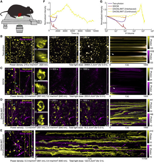
Figure 4. DAOSLIMIT facilitates long-term, high-speed subcellular imaging in mice (A) Illustrations of the imaging schematic. (B–D) Orthogonal MIPs (marked by the yellow dashed lines) of the neutrophil (yellow, Ly-6G/Ly-6C) and blood vessels (magenta, WGA) at different time stamps, imaged by high-speed two-photon microscopy (B), SDCM (C), and DAOSLIMIT with interleaved exposure (D). The kymograph with MIP along yt plane shows the bleaching process. Both the power densities during excitation and the total light doses are marked at the bottom. All the methods captured ∼150 two-color volumes. (E) Orthogonal MIPs and the kymograph obtained by DAOSLIMIT with continuous exposure at 3 volume/s for over 3 h, corresponding to 17,550 two-color volumes. (F and G) Curves of the normalized average intensity versus time (F) and the number of volumes (G) for the yellow channels. Scale bar, 10 μm. See also Table S1 and Video S2. |
Reprinted from Cell, 184, Wu, J., Lu, Z., Jiang, D., Guo, Y., Qiao, H., Zhang, Y., Zhu, T., Cai, Y., Zhang, X., Zhanghao, K., Xie, H., Yan, T., Zhang, G., Li, X., Jiang, Z., Lin, X., Fang, L., Zhou, B., Xi, P., Fan, J., Yu, L., Dai, Q., Iterative tomography with digital adaptive optics permits hour-long intravital observation of 3D subcellular dynamics at millisecond scale, 3318-3332.e17, Copyright (2021) with permission from Elsevier. Full text @ Cell