|
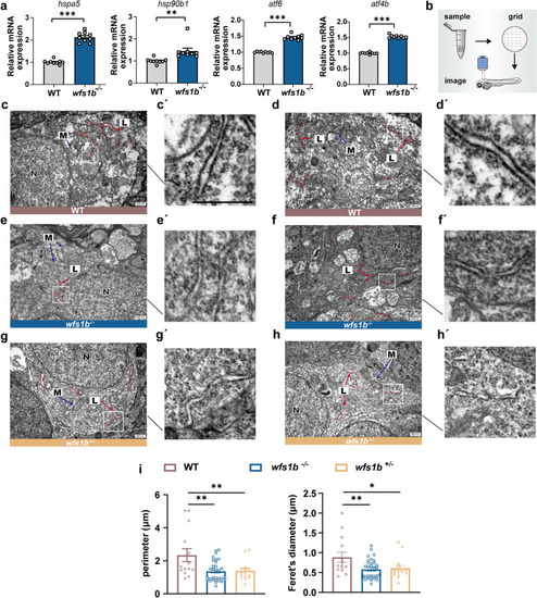
Loss of Wfs1b provokes the ER stress response. a qPCR analyses of up-regulated downstream genes of the UPR pathway. hspa5, P < 0.0001; hsp90b1, P = 0.0046; atf6, P < 0.0001; atf4b, P < 0.0001. Assessed by unpaired t test. b Diagram of the electron microscope’s operation. c–h Representative TEM imaging showed the ER morphology in wildtype (upper), wfs1b−/− mutant (middle) and wfs1b± mutant (bottom) larvae brain, indicating that the ER morphology was ruptured. (C′-H′) were the ER ultrastructure under magnification from the white boxes in c–f. Scale bar, 500 nm. L, rough endoplasmic reticulum; N, cell nuclei; M, mitochondria. i Statistical diagram of perimeter and Feret’ diameter among the wildtype, the wfs1b−/− and wfs1b+/- mutant zebrafish larvae. Assessed by ordinary one-way ANOVA
|