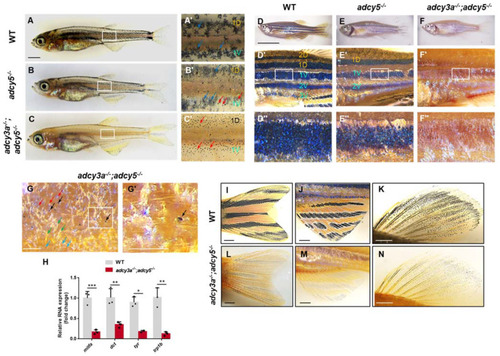
adcy3a and adcy5 are required for adult melanin patterning. (A–C) WT zebrafish develop metamorphic melanocyte stripes around 30 days, including 1D and 1V, along with corolla-like patterns of melanosomes in most melanocytes (A). adcy5-/- mutants establish normal metamorphic stripes, but some melanocytes present punctate distribution of melanosomes (B). adcy3a-/-;adcy5-/- double mutants are unable to form corolla distribution of melanosomes and instead punctate melanosomes (C). (A′–C′) are the magnified images of boxes in (A–C) (the red arrows indicate punctate melanocytes; the blue arrows indicate corolla melanocytes). Scale bar = 1000 μm in (A–C). (D–F) Adult wild-type zebrafish form five melanocyte stripes, including three ventral black stripes 1V, 2V, and 3V, and two dorsal stripes 1D and 2D along the dorsoventral axis (D). adcy5-/- mutants (E) show three melanocyte stripes, including black stripes 1D, 1V, and 2V, with a lower density of melanocytes, compare to WT zebrafish (F). adcy3a-/-;adcy5-/- double mutants fail to establish normal melanocyte stripes, along with a few punctate melanocytes (F). (D′–F′) are the partially magnified images of (D–F), and (D″–F″) are the magnified images of boxes in (D′–F′). 1D: dorsal primary black stripe; 1V: ventral primary black stripe; 2D: dorsal secondary black stripe; 2V: ventral secondary black stripe; 3V: the third ventral black stripe. Scale bar = 10 mm in (D–F). (G) Adult adcy3a-/-;adcy5-/- double mutants develop a few pigmented melanocytes, along with many unpigmented melanocytes (the red arrows indicate unpigmented melanocytes; the black arrows indicate pigmented melanocytes; the blue arrows indicate xanthophores; the green arrows indicate iridophores). (G′) is the partially magnified images of boxes in (G). Scale bar = 100 μm. (H) qPCR analyses of mitfa, dct, tyr, and trp1b expression in the adult skin from WT zebrafish or adcy3a-/-;adcy5-/- mutants (Student’s t-test, * p < 0.05, ** p < 0.01, *** p < 0.001). (I–N) The caudal, anal, and dorsal fins in adcy3a-/-;adcy5-/- double mutants show highly aggregated melanosomes compared with those in WT zebrafish. Scale bar = 1000 μm.