
Figure 4
- ID
- ZDB-FIG-221214-280
- Publication
- Zhang et al., 2022 - Requirement of Zebrafish Adcy3a and Adcy5 in Melanosome Dispersion and Melanocyte Stripe Formation
- Other Figures
- All Figure Page
- Back to All Figure Page
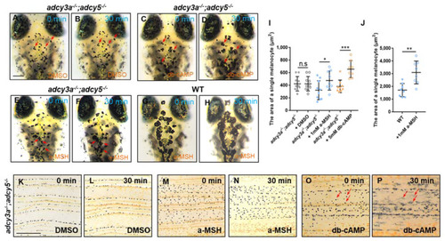
Activation of PKA ameliorates melanosome aggregation in adcy3a-/-;adcy5-/- double mutants. (A–H) 10-day-old adcy3a-/-;adcy5-/- double mutants have no response to DMSO treatment (A,B), but show significant dispersion of melanosomes when exposed to db-cAMP for 30 min (C,D). The melanosomes in a few melanocytes show mild dispersion when exposed adcy3a-/-;adcy5-/- double mutants to a-MSH for 30 min (E,F), while they dramatically disperse in almost all melanocytes in WT larvae (G,H). The red arrows indicate the same melanocytes from the same embryo before and after treatment with chemical compounds. Scale bar = 250 μm in (A–H). (I,J) Statistical analyses of the pigmented area of single melanocyte in adcy3a-/-;adcy5-/- double mutants (I) and WT embryos (J) before and after treatment with DMSO, a-MSH, and db-cAMP, respectively (Student’s t-test n.s., not significant, p > 0.05, * p < 0.05. ** p < 0.01. *** p < 0.001). (K–P) No melanosome dispersion is detected in DMSO (K,L) or a-MSH-treated caudal fins (M,N) derive from adcy3a-/-;adcy5-/- double mutants, but almost all melanosomes disperse in db-cAMP-treated mutant caudal fins (O,P). The red arrows indicate some dramatically dispersed melanosomes from the same caudal fin before and after treatment with db-cAMP. Scale bar = 250 μm in (K–P). |
| Fish: | |
|---|---|
| Conditions: | |
| Observed In: | |
| Stage Range: | Days 7-13 to Adult |