
FIGURE
Fig. 8
Fig. 8
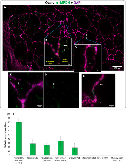
Fig. 8. IMPDH forms cytoophidia in the ovary of adult fish. Immunofluorescence images of the ovarian tissue of adult zebrafish cryosections for IMPDH (green) and DAPI (magenta). (A–E) Cytoophidia were observed in the vitellogenic and pre-ovulatory follicles. Cytoophidia were frequently located in the thick zona radiata of oocytes (B). Arrows indicate the cytoophidia in (B–E). Scale bars = 100 μm in (A); 10 μm in (B–E). (F) Quantification of cells presenting cytoophidia in the different tissues of adult fish. (n) indicate the number of cells counted. Error bars = S.D. |
Expression Data
| Antibody: | |
|---|---|
| Fish: | |
| Anatomical Terms: | |
| Stage: | Adult |
Expression Detail
Antibody Labeling
Phenotype Data
Phenotype Detail
Acknowledgments
This image is the copyrighted work of the attributed author or publisher, and
ZFIN has permission only to display this image to its users.
Additional permissions should be obtained from the applicable author or publisher of the image.
Reprinted from Developmental Biology, 478, Keppeke, G.D., Chang, C.C., Antos, C.L., Peng, M., Sung, L.Y., Coelho Andrade, L.E., Liu, J.L., IMPDH forms the cytoophidium in zebrafish, 89-101, Copyright (2021) with permission from Elsevier. Full text @ Dev. Biol.