
Fig. 6
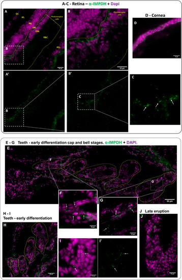
Fig. 6. IMPDH forms cytoophidia in the retina and teeth of adult fish. Immunofluorescence images of retina and teeth of adult zebrafish cryosections for IMPDH (green) and DAPI (magenta). (A–D) Abundant cytoophidia were observed in the outer nuclear layer and outer plexiform layer of the retina (arrows in C), and some were found in the rods and cones layer (A–C), but no cytoophidia were detected in the cornea (D). Highlighted by yellow dashed lines in (A″ and B″) is the region where photoreceptor cells are located. (B) Magnified image of selected region in (A). (C) Magnified image of selected region in (B). GC = ganglion cells, IPL = inner plexiform layer, INL = inner nuclear layer, OPL = outer plexiform layer, ONL = outer nuclear layer, R&C = rods and cones, PCL = pigment cell layer. (E–J) Cytoophidia were present in cells in the interior of teeth at early differentiation cap and bell stages (highlighted by yellow dashed lines in E and H), but not in the late eruption phase (J). (F and G) Magnified image of selected region in (E). (I) Magnified image of selected region in (H). Arrows in F, G and I indicate cytoophidia. Scale bars = 50 μm in (E); 20 μm in (A) and (H); 10 μm in (B), (D), (F), (G), (I) and (J). |
| Antibody: | |
|---|---|
| Fish: | |
| Anatomical Terms: | |
| Stage: | Adult |
Reprinted from Developmental Biology, 478, Keppeke, G.D., Chang, C.C., Antos, C.L., Peng, M., Sung, L.Y., Coelho Andrade, L.E., Liu, J.L., IMPDH forms the cytoophidium in zebrafish, 89-101, Copyright (2021) with permission from Elsevier. Full text @ Dev. Biol.