
Fig. 4
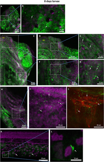
Whole-mount immunofluorescence of 8dpf larvae with anti-IMPDH2 antibody (shown in green) and DAPI (magenta). (A) Cytoophidia in the eye. (B) Zoom-in view of selected region in (A). (C) Ventral view of the head of larva. (D) Many cytoophidia were observed in the Meckel's cartilage (highlighted by yellow dashed lines in (C) and (D). (E) Zoom-in view of selected region in (D). (F) Cytoophidia were observed in the operculum. (G) Magnified image of selected region in (F). (H) Ventral view of ceratobranchials. (I and J) Zoom-in view of selected area in (H). The auto-fluorescence (red) shown in (J) reveals the contour of pharyngeal teeth, with abundant cytoophidia. (K and L) Some cells in the fin display cytoophidia. Arrows in all panels indicate cytoophidia. Scale bars = 50 μm in (A), (C), (H) and (K); 20 μm in (D), (F), (I) and (J); 10 μm in (B), (E), (G) and (L). |
| Antibody: | |
|---|---|
| Fish: | |
| Anatomical Terms: | |
| Stage: | Days 7-13 |
Reprinted from Developmental Biology, 478, Keppeke, G.D., Chang, C.C., Antos, C.L., Peng, M., Sung, L.Y., Coelho Andrade, L.E., Liu, J.L., IMPDH forms the cytoophidium in zebrafish, 89-101, Copyright (2021) with permission from Elsevier. Full text @ Dev. Biol.