
FIGURE
Fig. 5
Fig. 5
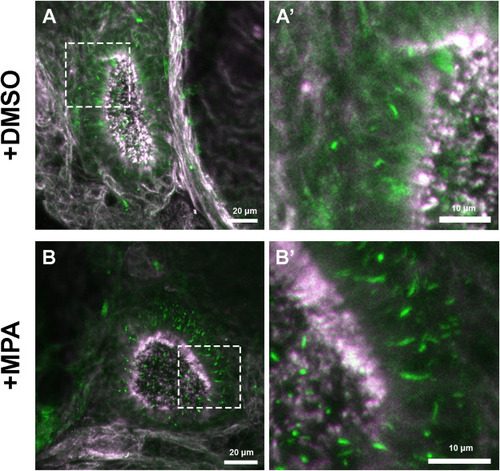
Fig. 5. MPA promotes IMPDH cytoophidium assembly in olfactory organ of larvae. Immunofluorescence images of the olfactory organ of 8 dpf larvae treated with 0.1% DMSO (A) or 100 μM MPA (B) for 1 day. (A′ and B′) Magnified images of selected areas in (A) and (B). IMPDH is shown in green and tubulin is shown in magenta. Scale bars = 20 μm in (A) and (B); 10 μm in (A′) and (B′). |
Expression Data
| Antibody: | |
|---|---|
| Fish: | |
| Condition: | |
| Anatomical Term: | |
| Stage: | Days 7-13 |
Expression Detail
Antibody Labeling
Phenotype Data
| Fish: | |
|---|---|
| Condition: | |
| Observed In: | |
| Stage: | Days 7-13 |
Phenotype Detail
Acknowledgments
This image is the copyrighted work of the attributed author or publisher, and
ZFIN has permission only to display this image to its users.
Additional permissions should be obtained from the applicable author or publisher of the image.
Reprinted from Developmental Biology, 478, Keppeke, G.D., Chang, C.C., Antos, C.L., Peng, M., Sung, L.Y., Coelho Andrade, L.E., Liu, J.L., IMPDH forms the cytoophidium in zebrafish, 89-101, Copyright (2021) with permission from Elsevier. Full text @ Dev. Biol.