
Fig. 2
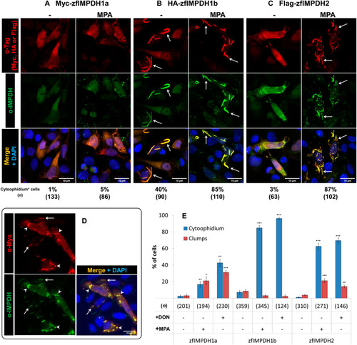
Fig. 2. Diverse zfIMPDHs cytoophidium-forming properties in vitro. (A–C) zfIMPDH isoforms were cloned with N-terminal tags and expressed in HeLa cells. Immunofluorescence probing on transfected HeLa cells under conditions with or without treatment of MPA (100 μM for 2 h). Myc-tag, HA-tag and Flag-tag are shown in red and antibody against IMPDH is shown in green. Cytoophidia formed by zfIMPDH are indicated by arrows in B and C. The proportion of cells with cytoophidia and the number of cells were counted (n) is shown below each panel. (D) HEp-2 cells expressing zfIMPDH1a were treated with DON (100 μM for 2 h) before fixation. Cytoophidia and clumps that contain zfIMPDH1a are indicated by arrows and arrowheads, respectively. Scale bars = 10 μm. (E) Bar graph showing the quantification of the proportion of zfIMPDH expressing HEp-2 cells with cytoophidia or clumps. Only transfected cells were counted and the number of cells counted in each group is shown as (n). (Error bars = S.E.M. ∗p < 0.05; ∗∗p < 0.01; ∗∗∗p < 0.001, proportions were compared by t-test with the respective isoform without treatment.) In all groups with MPA treatment, endogenous cytoophidia in non-transfected cells are labeled by the α-IMPDH antibody. |
Reprinted from Developmental Biology, 478, Keppeke, G.D., Chang, C.C., Antos, C.L., Peng, M., Sung, L.Y., Coelho Andrade, L.E., Liu, J.L., IMPDH forms the cytoophidium in zebrafish, 89-101, Copyright (2021) with permission from Elsevier. Full text @ Dev. Biol.