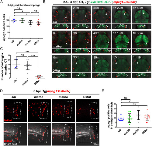
Microglia colonization of the optic tectum is defective in DMut. (A) Quantification of peripheral macrophages in 3 dpf siblings (n = 7), mafba mutants (n = 7), mafbb mutants (n = 3), and DMut (n = 7) in Tg(mpeg1:DsRedx) background (mean ± SD; Student’s t test; nonsignificant [ns] P > 0.05, *P < 0.05). (B) Coronal and transverse views of time-lapse imaging pictures of the midbrain of siblings, mafba mutants, and DMut in Tg(-2.8elavl3:eGFP;mpeg1:DsRedx) transgenic background where microglia are labeled in red and neurons are marked in green. Dashed lines indicate the optic tectum (OT) region. White arrows indicate microglia that have entered the OT. (C) Quantification of microglia number entering the OT in siblings (n = 3), mafba mutants (n = 8), and DMut (n = 5) from 2.5 to 3 dpf (mean ± SD; Student’s t test; nonsignificant [ns] P > 0.05, ***P < 0.001). (D) Representative tail images of peripheral macrophages surrounding the injury sites 6 h post injury (hpi) in 3 dpf siblings, mafba mutants, mafbb mutants, and DMut in Tg(mpeg1:DsRedx) transgenic background. Macrophages are labeled in red color. (E) Quantification of peripheral macrophages surrounding the injury sites 6 h post injury in 3 dpf siblings (n = 11), mafba mutants (n = 8), mafbb mutants (n = 10), and DMut (n = 15) in Tg(mpeg1:DsRedx) transgenic background (mean ± SD; Student’s t test; nonsignificant [ns] P > 0.05, *P < 0.05).
|
