|
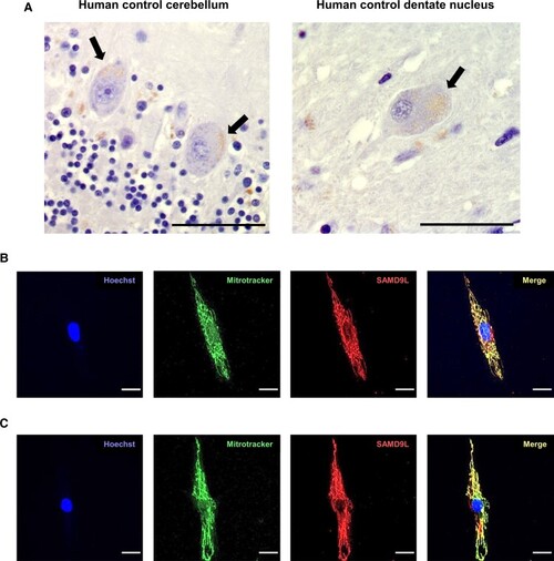
Mitochondrial localization of SAMD9L in human cerebellar Purkinje cells and multipolar neurons of the dentate nucleus and fibroblasts. (A) Immunostaining with anti-SAMD9L antibody in human control cerebellar sections revealed a punctate staining mainly in Purkinje cell soma (left) and in multipolar neurons (right) of the human dentate nucleus indicative of mitochondrial staining. Black arrows point to SAMD9L staining in Purkinje and multipolar neurons. (B and C) Immunofluorescence staining of fibroblasts from human control (B) and a M-SCA affected patient (C) showed co-localization of SAMD9L to mitoTracker Red CMXRos demonstrating mitochondrial co-localization. Magnification bars: 50 µm (A), 20 µm (B and C).
|