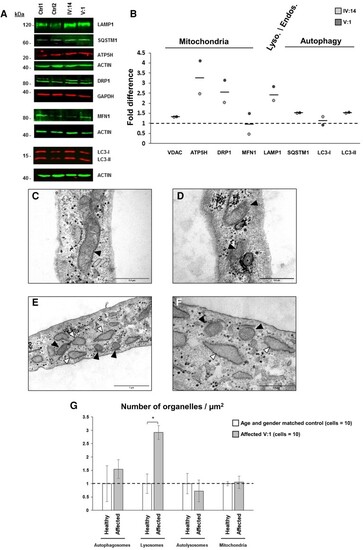
Mitochondrial and lysosome/autophagy alterations in fibroblasts from ataxia patients. (A and B) The ATP5H and DRP1 mitochondrial associated proteins levels were found increased without changes in VDAC and MFN1 suggestive of mitochondrial biogenesis. Furthermore, lysosomal LAMP1 together with SQSTM1 and LC3-II protein levels were found increased suggestive of mitophagy. LAMP1, MFN1, SQSTM1, ATP5H AND LC3 proteins levels were normalized to actin B (ACTIN) and DRP1 proteins levels to GAPDH (Supplementary Figs 22–27). Controls values were set to 1 (dotted line). TEM showed diffused crests in the mitochondrial matrix (C) together with some mitophagy (D) and dilated endoplasmic reticulum (E and F) compared with age-matched control (Supplementary Fig. 13). (G) One-way ANOVA confirmed significantly increased number of lysosomes on patients’ fibroblasts samples (N = 10) compared with controls [N = 10; F(1,18) = 6.135, P = 0.023] (Supplementary Fig. 13). Black arrowheads point to mitochondria; white arrowheads point to the dilated endoplasmic reticulum. Asterisks indicate autophagosomes. Magnification bars: 0.5 µm (C, D and F), 1 µm (E). Asterisk denotes significance at P < 0.05. Vertical bars denote SEM.
|
