IMAGE

Figure 4

ID
ZDB-IMAGE-220324-23
Source
Figures for Corral-Juan et al., 2022
Image
Figure Caption

Figure 4

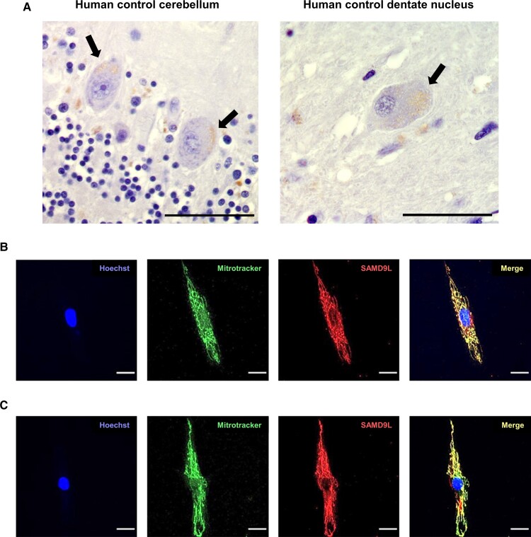
Mitochondrial localization of SAMD9L in human cerebellar Purkinje cells and multipolar neurons of the dentate nucleus and fibroblasts. (A) Immunostaining with anti-SAMD9L antibody in human control cerebellar sections revealed a punctate staining mainly in Purkinje cell soma (left) and in multipolar neurons (right) of the human dentate nucleus indicative of mitochondrial staining. Black arrows point to SAMD9L staining in Purkinje and multipolar neurons. (B and C) Immunofluorescence staining of fibroblasts from human control (B) and a M-SCA affected patient (C) showed co-localization of SAMD9L to mitoTracker Red CMXRos demonstrating mitochondrial co-localization. Magnification bars: 50 µm (A), 20 µm (B and C).

Acknowledgments
This image is the copyrighted work of the attributed author or publisher, and ZFIN has permission only to display this image to its users. Additional permissions should be obtained from the applicable author or publisher of the image. Full text @ Brain Commun