
Figure 2
- ID
- ZDB-FIG-220104-139
- Publication
- Li et al., 2021 - The Adaptor Protein Lurap1 Is Required for Cell Cohesion during Epiboly Movement in Zebrafish
- Other Figures
- All Figure Page
- Back to All Figure Page
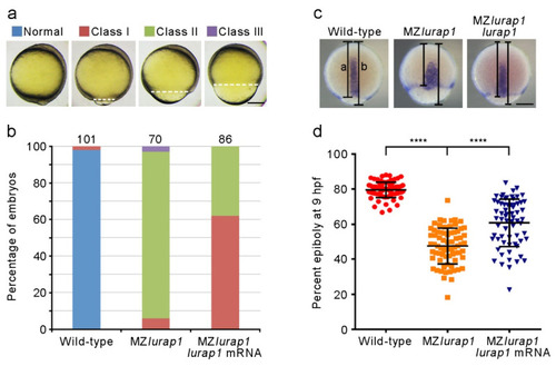
Rescue of epiboly delay in MZlurap1 mutant embryos by Lurap1. (a) Classification of the severity of epiboly delay at 10 hpf: normal embryos complete epiboly; class I embryos reach 90% epiboly; class II embryos show 75% to 90% epiboly; class III embryos fail to accomplish 75% epiboly. Broken lines indicate the leading edge of the blastoderm margin. Scale bar: 200 µm. (b) Graph shows the proportion of embryos with different degrees of epiboly delay. Numbers on the top of each stacked column represent total embryos scored from three independent experiments, and color keys are as indicated in (a). The results were analyzed independently in a blended manner. (c) Determination of the rescue efficiency by in situ hybridization analysis of tbxta to visualize the leading edge of the blastoderm margin at 9 hpf. Epiboly progression was calculated as the distance from the animal pole to the edge of the blastoderm margin (a) relative to the length of the animal-vegetal axis (b). Scale bar: 200 µm. (d) Scatter plot shows the rescue of epiboly progression in MZlurap1 mutant embryos by wild-type lurap1 mRNA. Data are mean ± s.d. from three independent batches of embryos (****, p < 0.0001). |