
Fig. 3
- ID
- ZDB-FIG-211206-4
- Publication
- Jin et al., 2021 - Copper ions impair zebrafish skeletal myofibrillogenesis via epigenetic regulation
- Other Figures
- All Figure Page
- Back to All Figure Page
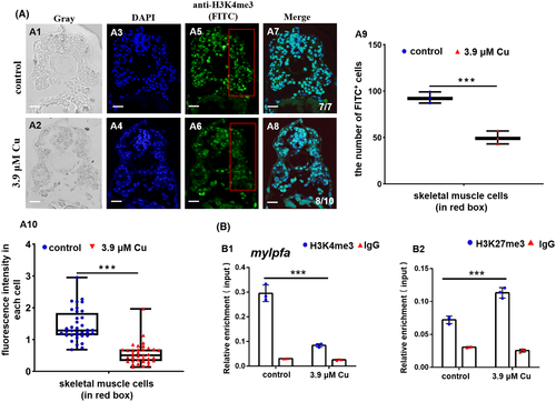
Cu2+-stressed zebrafish embryonic muscle cells exhibited reduction in both the H3K4me3 protein level and the binding enrichment of H3K4me3 protein on muscle fiber gene mylpfa promoter. A, Protein level of H3K4me3 in Cu2+-stressed zebrafish embryonic muscle cells. A1, A2, gray; A3, A4, DAPI; A5, A6, FITC; A7, A8, merged. A9, DAPI+ FITC+ cell numbers calculated in the fixed red boxes in A5 and A6, respectively. A10: FITC intensity calculated for each cell (50-100 cells/sample) in A5 and A6, respectively. Each dot represents the fluorescence intensity in each cell. B, Reduced binding enrichment of protein H3K4me3 (B1) while increased binding enrichment of protein H3K27me3 (B2) for the muscle fiber gene mylpfa promoter as revealed by chromatin immunoprecipitation assays (ChIP). Anti-H3K4me3 and Anti-H3K27me3 were used for ChIP assays in the control and the Cu2+-stressed embryonic cells, with anti-IgG used as negative control. Each experiment was repeated two or three times with similar results, and a representative result is shown. A1-A8, transverse sections. *P < .05, **P < .01, ***P < .001. NS, not significant. A1-A8, scale bar, 100 μm |