
Figure 2
- ID
- ZDB-FIG-211201-173
- Publication
- Chen et al., 2021 - Knockout of mafba Causes Inner-Ear Developmental Defects in Zebrafish via the Impairment of Proliferation and Differentiation of Ionocyte Progenitor Cells
- Other Figures
- All Figure Page
- Back to All Figure Page
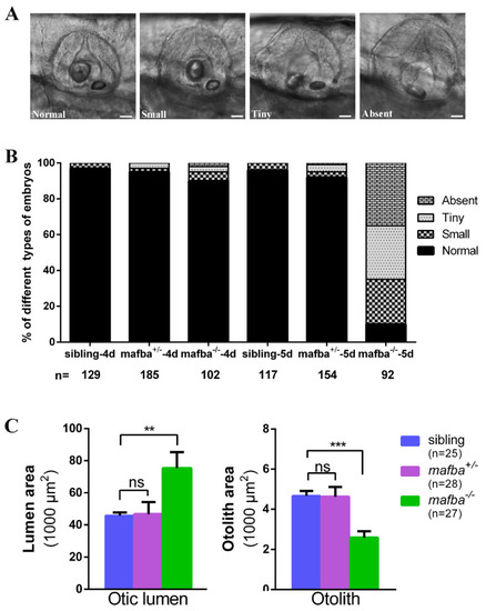
Deletion of mafba leads to inner-ear morphological defects. (A) The variable otolith sizes of mafba−/− mutant embryos at 5 dpf. According to the otolith sizes, mutant embryos were classified into four groups: normal, small, tiny, and absent. Scale bars: 40 µm. (B) Percentages of embryos in sibling and mafba−/− group at 4 dpf and 5 dpf; n, the number of observed embryos. (C) Statistical analysis of the otic lumen area and otolith area in different types of embryos at 5 dpf. Individual embryos were randomly picked from each type for statistical analysis. The otic lumen and otolith areas were measured with SPOT Advanced software (version 4.6) in the focal plane representing the maximal area. Data are represented as mean ± SD; ns, p > 0.05; **, p < 0.01; ***, p < 0.001. |
| Fish: | |
|---|---|
| Observed In: | |
| Stage Range: | Day 4 to Day 5 |