|
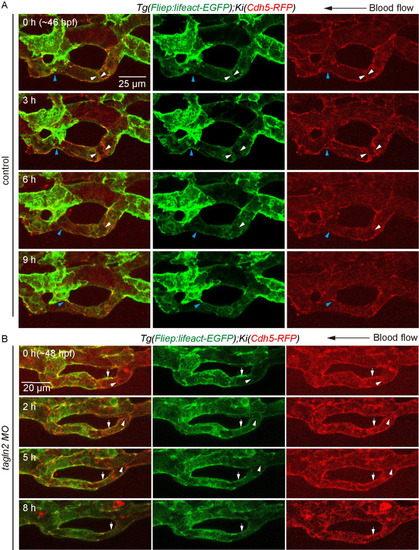
Knockdown of <italic toggle='yes'>tagln2</italic> impairs junction remodeling and rearrangement of F-actin cytoskeleton in CV pruning.(A) and (B) Time-lapse live imaging of junction remodeling and actin cytoskeleton dynamics in CV prunig in Tg(Fliep:Lifeact-EGFP);KI(cdh5-mRFP) embryos. (A) Control embryo shows a detachment of junction (blue arrowheads) and an opposite direction of junction movement (white arrowheads) as multicellular tube became stenosis. F-actin forms at cdh5-positive junction (white and blue arrowheads), and undergoes a similar rearrangement as cdh5-positve junctions. Seven vascular loops are taken time-lapse live imaging at the stage of multicellular tube. Scale bar: 25 μm. (B) In tagln2 morphant, cdh5-positive junction moves to the right and connected with the adjacent one (white arrowheads), and F-actin undergoes similar rearrangement within it (white arrowheads). However, depolymerization of F-actin is also observed at junctions (white arrowheads). Six vascular loops are taken time-lapse live imaging. Scale bar: 20 μm.
|