|
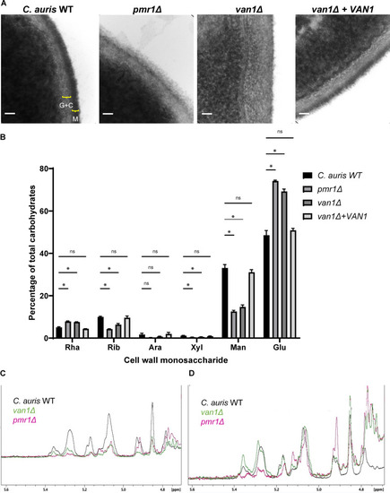
C. aurispmr1Δ and van1Δ strains display an altered cell wall structure that contains less mannan. (A) C. auris yeast were imaged via transmission electron microscopy at 383,000× magnification, and scale bars represent 50 nm. Brackets denote distinct cell wall layers: G+C, β-glucan and chitin; M, mannan. (B) The monosaccharide compositions of cell walls were measured by gas chromatography, n = 5, mean with SEM shown, *, P < 0.05; ns, not significant by one-way ANOVA with Holm-Sidak multiple comparisons to C. auris WT. Rha, rhamnose; Rib, ribose; Ara, arabinose; Xyl, xylose; Man, mannose; Glu, glucose. (C and D) The structures of isolated mannans were analyzed by 1H NMR and COSY spectra. C shows the 1H NMR spectra for each strain following mannan isolation. In panel D, the intensities were adjusted to the resonance assigned to sidechain-linked backbone α1-6-linked mannosyl repeat units (5.07 ppm) to compare mannan structures.
|