|
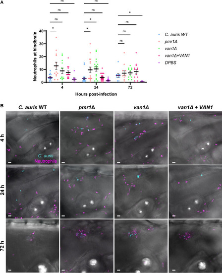
C. auris mannan mutants stimulate increased neutrophil recruitment in the larval zebrafish hindbrain. C. auris strains were injected into the hindbrains of larvae from a cross between the Tg(lyzC:RFP) and Tg(mpeg:GFP) lines at 2 days postfertilization. Fluorescence microscopy was utilized to measure recruitment of neutrophils to the hindbrain at 4, 24, and 72 h postejection. (A) At each time point, fluorescent neutrophils were manually enumerated from maximum intensity projections from z-stacks; n = 9 to 23, experiments were performed in three replicates; the mean with SEM are shown; *, P < 0.05; ns, not significant by Brown-Forsythe and Welch ANOVA with Dunnett’s T3 multiple comparisons to C. auris WT. (B) Representative fluorescence microscopy images of neutrophil recruitment to zebrafish hindbrain are shown (magenta = neutrophils, cyan = C. auris cells). Scale bar = 20 μm.
|