|
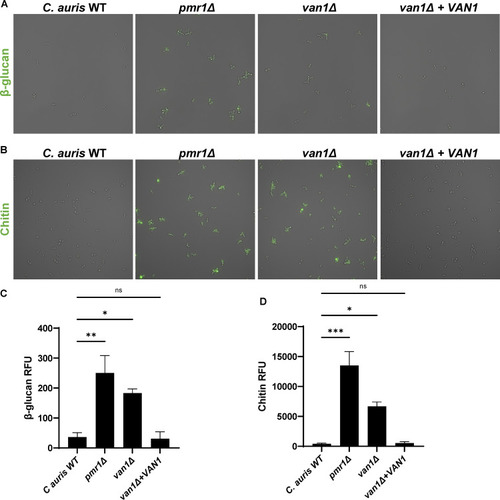
C. aurispmr1Δ and van1Δ strains display increased cell surface PAMPs. (A) Cell surface β-glucan was labeled using Fc:dectin-1 protein with Alexa Fluor 488-conjugated anti-human IgG Fc antibody and imaged by fluorescence microscopy. (B) Cell surface chitin was labeled with wheat germ agglutinin conjugated to fluorescein isothiocyanate (WGA-FITC) and assessed by fluorescence microscopy. (C and D) Total surface β-glucan and chitin were quantified by plate reader measurements of fluorescence, n = 3 mean with SEM shown, *, P < 0.05; **, P < 0.01; ***, P < 0.001 by one-way ANOVA with Holm-Sidak multiple comparisons to C. auris WT; ns, not significant.
|