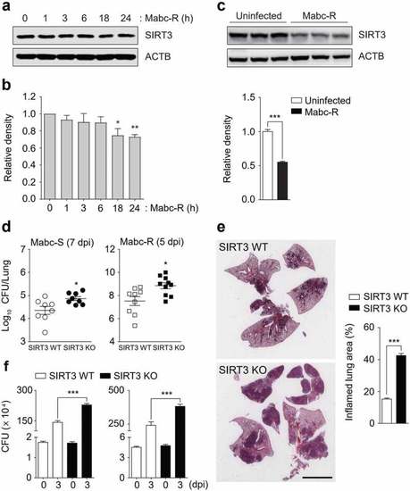
SIRT3 is essential for host defense against mycobacterial infection in vivo and in vitro. (a and b) WT BMDMs were infected with Mabc-R (MOI = 3) at the indicated times. Actin protein levels were evaluated by immunoblotting as an internal control. (b) Quantification of results on (a). (c) Western blot analysis of the lung tissues from SIRT3 WT and KO mice left uninfected or infected intranasally with Mabc-R (1 × 10⁷ CFU) for 3 days. Quantifiation of results on top. (d) SIRT3 WT and KO mice were infected intranasally with various CFUs of Mabc-R (1 × 10⁷ CFU) or Mabc-S (1 × 10⁷ CFU) and monitored at 5 or 7 days post-infection (dpi). Data are shown as log pulmonary CFU. (e) Lung histopathology by H&E staining of SIRT3 WT and KO mice infected with Mabc-R for 5 days. Right, Quantification of results on left. Scale bar, 5 mm. (f) Intracellular survival of Mabc-S assessed by a CFU assay. SIRT3 WT and KO BMDMs were infected with Mabc-S (MOI = 1, for left; MOI = 3, for right) for 4 h, and then lysed to determine intracellular bacterial loads at 0 and 3 dpi. *P < 0.05, **P < 0.01, ***P < 0.001. Non-parametric test (b and d); Student’s t-test (c below and e right); One-way ANOVA (f). Data represent three independent experiments (a, c top, and e left), and values represent means (± SEM) from three or four independent experiments performed in triplicate (b, c bottom, e right, and f).
|
