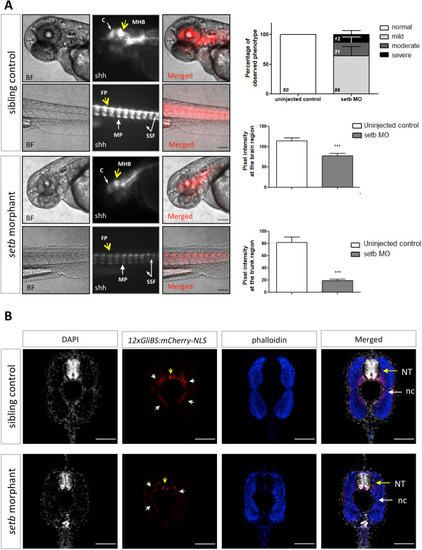
Activation of Hh signaling is reduced in setb morphants. (A) Fluorescent and brightfield images of 48 hpf setb-morpholino injected and control sibling Tg12x_Gli embryos. Embryos are placed anterior to left, dorsal up. Scale bars 100 μm. Quantification of pixel intensity at 48 hpf of the morphologically mildly affected embryos (***p < 0.001). Data are expressed as mean ± SEM (n = 129, after exclusion of the injected embryos developing apoptotic tissue at 24 hpf). Percentage of phenotypic scoring of 48 hpf morphants is presented with mean ± SEM and the numbers of total embryos per group is indicated. At the brain region: cerebellum (c, white arrow), mid-hindbrain boundary (MHB, yellow arrow). At the trunk region: floor plate (FP, yellow arrow), slow muscle pioneer cells (MP, white arrow, high mCherry signal), superficial slow fibers (SSF, white arrows, mid mCherry signal). (B) Confocal images of cross-sections throughout the trunk region of 48 hpf setb morphants and age-matched siblings. Note the reduction of mCherry+ cells at the ventral side of neural tube (yellow arrows) and at the slow fibers surrounding notochord (white arrows) of setb morphants; indication of lower activation levels of the Hh pathway. Sections were stained with phalloidin-633 (blue) and DAPI (grey). NT: neural tube, nc: notochord. Scale bars 50 μm.
|
