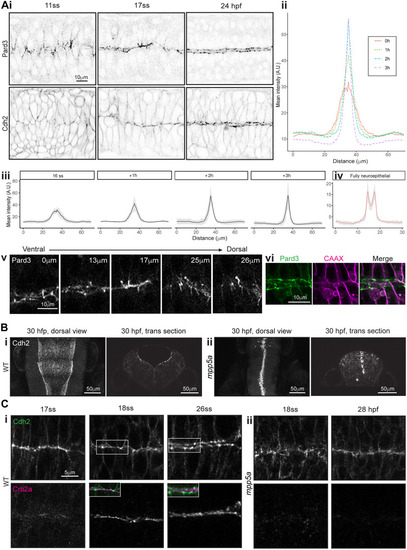
Mpp5a-dependent remodelling of midline adhesions. (Ai) Images from confocal time-lapse movies of Pard3-EGFP and Cdh2-GFP embryos in horizontal orientation at 11-somite stage (ss), 17ss and 24 hpf stages. Comparable images were seen from three embryos from each transgenic line. (Aii,Aiii) Mean intensity profiles from six embryos, quantifying Pard3 intensity across the basal-to-basal width of the developing neuroepithelium over time, starting at the 16-somite stage. Standard deviation is shown as a grey ribbon around the line profile for each time point in Aiii. (Aiv) Mean intensity profiles from the same six embryos, quantifying Pard3 intensity across the basal-to-basal width of the neuroepithelium at the fully neuroepithelial stage. (Av) Horizontal confocal planes of 17-somite stage neural rod showing Pard3-EGFP expression at five different dorsoventral levels. The single elevated plane of expression at the left-right interface, seen at the level of 17 μm, lies dorsal to levels where apical rings are already formed and ventral to levels where expression is more prominent in mediolateral streaks. (Avi) Single horizontal plane confocal section of Pard3-EGFP and mNeptune2.5-CAAX and merge at ∼24 hpf. Plasma membranes meet at the tissue midline, and Pard3 is now largely located in two parallel parasagittal domains. (B) Horizontal and transverse confocal sections of 30 hpf. Wild-type (WT; Bi) and mpp5am227 mutant (Bii) Cdh2-GFP embryos at the hindbrain level. The hindbrain lumen remained closed in five of five mpp5am227 mutant embryos and is always open in wild types. (C) Horizontal confocal sections of wild-type (Ci) and mpp5am227 mutant (Cii) Cdh2-GFP embryos at the anterior spinal cord level, stained for Crb2a. Insets in the 18- and 26-somite stages of wild types show merged images of Crb2a and Cdh2-GFP expression. (Ci) In wild-type embryos, Cdh2 and Crb2a were colocalised at the midline at the 18-somite stage (four of four embryos), but Cdh2-GFP was displaced basolaterally to form two independent stripes of expression either side of the midline by the 26-somite stage (eight of eight embryos). In mpp5am227 mutants, Crb2a was not present at the midline at the 18-somite stage (four of four embryos), and Cdh2-GFP remained in a single expression domain at the tissue midline even as late as 28 hpf (five of five embryos).
|