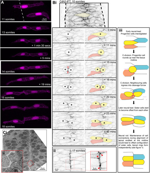
Sister cells remain attached via their corners. (A) Images from time-lapse movie projection in dorsal orientation of mosaically labelled neuroepithelial cells in the hindbrain of an 11-somite-stage wild-type embryo. Membrane and nuclei are labelled in magenta. By the 13-somite stage, both cells had undergone C-division, resulting in pairs of sister cells attached across the tissue midline. The top cell pair was followed over time until the 18-somite neural rod stage, when both cell pairs were imaged. The configuration of cell pair connections was assessed from several different experiments at the neural rod stage (∼16-18 somites), and 26 of 31 pairs of cells from five embryos at neural rod stages were found clearly to be attached via their corners. The remaining five were either connected via a more ‘en face’ configuration or their configuration was uncertain (e.g. owing to a very thin connection point). (Bi) Single z-planes from a time-lapse movie of a C-division (yellow cell) starting at the 10-somite stage (neural keel), from a Cdh2-tFT transgenic embryo. The image contrast was increased in the reference image at the top to highlight that Cdh2 was concentrated at the interdigitation zone between cells around the tissue midline at 0 min. Cdh2-GFP becomes strongly concentrated in the cleavage furrow (time point 15 min), and neighbouring cells ingress into the cleavage furrow (21 of 21 divisions, red arrows). In this example, the pink cell that ingresses into the cleavage furrow gains a contralateral contact with the contralateral daughter of the C-division. As a result of this contact, the contralateral daughter (yellow cell on right) becomes attached to two contralateral cells; one is its sister cell from the C-division and the other is one of its sister's neighbouring cells (the pink cell in this example). (Bii) 5 µm projection of 17-somite stage neural rod from a Cdh2-tFT transgenic embryo. Endfeet are aligned along a centrally located midline. Cdh2-GFP is upregulated along the midline, particularly at cell corners (red arrowheads in magnified region). (Biii) Model depicting the co-ingression of neighbouring cells into the cleavage furrow during C-division (yellow cells) and the subsequent offsetting of sister cells from each other, which precedes apical ring formation, based on images similar to those in Bi. Red lines and dots represent high levels of Cdh2 associated with the dividing cell. The ingression of either ipsilateral (e.g. orange) or contralateral (e.g. blue) neighbours into the cleavage furrow promotes the formation of multiple contralateral connections across the midline. For example, the left-hand yellow sister cell becomes attached to both its right-hand yellow sister cell and the ingressing blue cell. The right-hand yellow sister cell becomes attached to both its left-hand yellow sister cell and the ingressing orange cell. (Ci) Transmission electron micrographs of a 20 hpf embryo hindbrain in transverse orientation. The lumen has started to open from the midline. The interface between contralateral cells has a striking ‘zig-zag’ pattern (three of three 19-20 hpf embryos). (Cii) Inset magnified region from Ci.
|