|
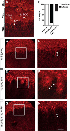
Knockdown of <italic>mondoa</italic> affects microtubule stability.(A) Confocal image of the blastoderm margin of an uninjected embryo at sphere stage; microtubules are stained with a mouse α-tubulin antibody followed by an anti-mouse Alexa Fluor 488-coupled antibody. DEL; deep cell layer; MT, microtubules; MTOC, microtubule organization center; YCL, yolk cytoplasmic layer; YSL, yolk syncytial layer; YSN, yolk syncytial layer nuclei. (B) Quantification of effects on YSL microtubule organization in uninjected (n = 9), mondoa-mo injected (n = 16), and mondoa-mo injected + pregnenolone (P5) treated (n = 9) embryos. Presence of stellar structures as shown in (F) = ‘affected’, normal appearance as in (A) = ‘unaffected’. (C–H) Overview (C, E, G) and close-up (D, F, H) images of uninjected embryos (C, D), mondoa-mo morphants (E, F) and P5 treated mondoa-mo morphants (G, H). Arrowheads indicate long parallel MT in uninjected and P5 rescued embryos (D, H) and short MT asters in untreated morphants (F). Scale bar: 90 µm; for higher magnifications, 30 µm.
|