|
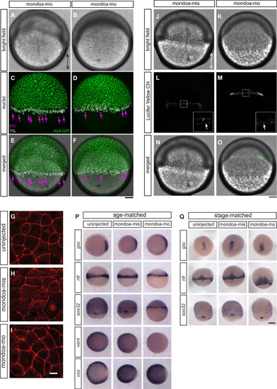
EVL epiboly and integrity, endocytic vesicle formation at the marginal rim and overall blastoderm patterning are not severely perturbed in <italic>mondoa</italic> morphants.(A–F) Tg(h2afva:h2afva-GFP) (H2A-GFP) transgenic embryos were injected with either mondoa-mis (A, C and E) or mondoa-mo (B, D and F) at the one-cell stage. Phenotypes of representative embryos among those analyzed (n = 2 for mondoa-mis and n = 9 for mondoa-mo) at seven hpf are shown. (A, B) Brightfield images. (C, D) GFP channel. The positions of EVL- and YSL-nuclei as identified by confocal reflection images are shown with magenta and white circles, respectively. The arrows (magenta) indicate EVL nuclei vegetal to the blastoderm margin. (E, F) Bright field images merged with H2A-GFP positive nuclei. Animal pole up. Scale bar: 100 µm. (G–I) Phalloidin stainings of uninjected (G), mondoa-mis injected (H) and mondoa-mo injected (I) embryos, view of the lateral EVL at around sphere stage. No differences in cell shape or staining integrity are apparent between the three conditions. Scale bar: 22 µm. (J–O) Embryos were injected with either mondoa-mis (J, L and N; n = 6) or mondoa-mo (K, M and O; n = 5). (J, K) Brightfield images. (L, M) Lucifer Yellow CH-positive endocytic vesicles were observed at the marginal rim in both mondoa-mis injected embryos (L) and mondoa-mo morphants (M). The inset at the bottom right is a magnified view of the region in the white rectangle. The arrow (white) indicates the endocytic vesicles. (N, O) Bright field images merged with Lucifer Yellow CH-positive vesicles (green). Animal pole up. Scale bar: (J–O) 100 µm; insets in L and M: 33 µm. (P, Q) WISH against different patterning markers in uninjected embryos and in embryos injected with mondoa-mis or mondoa-mo. Markers examined were: no tail (ntl; for mesoderm; Schulte-Merker et al., 1994), goosecoid (gsc; for dorsal mesendoderm/organizer; Stachel et al., 1993), SRY-box containing gene 32 (sox32; for endoderm; Dickmeis et al., 2001; Kikuchi et al., 2001), and the ventral markers ventral homeobox (vox) and ventral expressed homeobox (vent) (Melby et al., 2000). To control for pattern changes caused only by developmental delay, we either fixed the embryos all at the same time since fertilization (i.e., age-matched) or at the same stage regardless of the developmental time elapsed until the stage was reached (i.e., stage-matched; in this case the MO dose had to be lowered to allow for further development of the morphants). (P) gsc expression is slightly expanded in the morphants in age-matched embryos, and vent expression is strongly reduced. Expression of vox, which functions redundantly with vent (Imai et al., 2001), only shows a slightly larger gap in the dorsal area. The patterns of the other germ layer markers appear indistinguishable from the controls. Embryos were fixed when uninjected controls had reached shield stage (gsc, ntl, vox, vent) or 70% epiboly (sox32). (Q) In stage-matched embryos, only ntl expression shows a slightly broadened expression in the morphants, potentially indicating difficulties with convergence/extension movements, with no changes observed for the other markers examined. Thus, sox32 expression shows normal expression domains in endoderm precursors and forerunner cells. Stages were 60% epiboly (gsc, ntl) and 80% epiboly (sox32). Scale bar: 0.2 mm.
|