FIGURE

Fig 2

ID
ZDB-FIG-200610-23
Publication
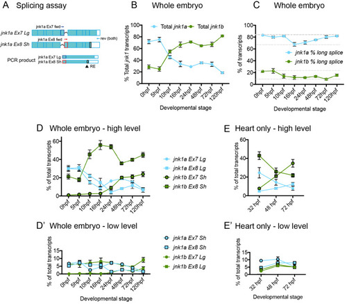
Santos-Ledo et al., 2020 - Alternative splicing of jnk1a in zebrafish determines first heart field ventricular cardiomyocyte numbers through modulation of hand2 expression
Other Figures
All Figure Page
Back to All Figure Page
Fig 2

Semi-quantitative <italic>jnk1a/b</italic> transcript assay.

(A) Schematic of assay for jnk1a (see methods for full details). RNA is extracted and a cDNA pool produced. PCR within linear phase performed using forward (fwd) primers specific for jnk1a/1b exons 7/8 and common reverse (rev) primers. The fraction with C-terminal extension is determined by restriction enzyme (RE) digestion. (B) Percentage of total jnk1a and jnk1b transcripts during embryonic development. 14 ss = 16 hpf. (C) Percentage of total Lg jnk1a and jnk1b transcripts during embryonic development. (D and D') Proportion of each individual alternatively spliced transcript during development in whole embryo. (E and E') Proportion of each individual transcript within isolated whole heart during development. jnk1a = blue, jnk1b = green, Ex7 = circle, Ex8 = square, Sh = outline black and Lg = no outline. n = 6–8 at all time points.

Expression Data
Genes:
Fish:
Anatomical Terms:
Stage Range: 1-cell to Day 5

Expression Detail
Antibody Labeling
Phenotype Data

Phenotype Detail
Acknowledgments
This image is the copyrighted work of the attributed author or publisher, and ZFIN has permission only to display this image to its users. Additional permissions should be obtained from the applicable author or publisher of the image. Full text @ PLoS Genet.