Figure 3—figure supplement 1

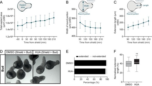
Cell proliferation is dispensable during blastoderm explant extension.

(A) Total blastoderm explant area prior and from shield stage onwards (−60 min: n = 24, N = 3; 0 min: n = 23, N = 3; 30 min: n = 23, N = 3; 60 min: n = 22, N = 3; 90 min: n = 21, N = 3; 120 min: n = 22, N = 3; 150 min: n = 23, N = 3; 180 min: n = 22, N = 3; 210 min: n = 23, N = 3). (B) Width of the round portion of blastoderm explants from shield stage onwards (90 min: n = 20, N = 3; 120 min: n = 22, N = 3; 150 min: n = 23, N = 3; 180 min: n = 22, N = 3; 210 min: n = 23, N = 3). (C) Length of the explant extension from shield stage onwards (90 min: n = 20, N = 3; 120 min: n = 22, N = 3; 150 min: n = 23, N = 3; 180 min: n = 22, N = 3; 210 min: n = 23, N = 3). Time in min. Schematic representation of the quantification method is shown at the top (A-C). (D) Bright-field single-plane images DMSO or HUA-treated blastoderm explants (treated from shield to bud stage; DMSO: n = 29, N = 3; HUA: n = 35, N = 3) at bud stage. Scale bar: 200 µm. (E) Percentage of extended or not-extended DMSO or HUA-treated blastoderm explants (treated from shield to bud stage; DMSO: n = 28, N = 3; HUA: n = 34, N = 3) at bud stage. (F) Normalized extension length of extended DMSO or HUA-treated blastoderm explants (treated from shield to bud stage; DMSO: n = 28, N = 3; HUA: n = 32, N = 3) at bud stage. ns, not significant (Unpaired t test).

Cell proliferation is dispensable during blastoderm explant extension.

Expression Data

Expression Detail
Antibody Labeling
Phenotype Data

Phenotype Detail
Acknowledgments
This image is the copyrighted work of the attributed author or publisher, and ZFIN has permission only to display this image to its users. Additional permissions should be obtained from the applicable author or publisher of the image. Full text @ Elife